Basal Cell Skin Cancer – Symptoms, Causes, Prevention
Basal cell skin cancer, clinically termed basal cell carcinoma (BCC), constitutes the most prevalent form of skin malignancy diagnosed in the United States, with dermatologists documenting over 3.6 million cases annually. This neoplasm arises from the uncontrolled proliferation of basaloid cells within the epidermis or hair follicles, typically manifesting decades after initial ultraviolet radiation damage occurs. Despite its status as the most common cancer overall, BCC maintains an exceptionally favorable prognosis when detected during early stages, characterized by indolent growth patterns and negligible metastatic potential.
The pathophysiology centers predominantly on cumulative DNA damage induced by ultraviolet B (UVB) radiation, though ultraviolet A (UVA) contributes indirectly through reactive oxygen species generation. Clinical presentations vary substantially among subtypes, ranging from nodular lesions with pearly translucence to superficial erythematous patches. While locally destructive capabilities necessitate prompt intervention, the condition’s mortality rate remains negligible compared to melanoma, reinforcing the importance of regular dermatological surveillance and photoprotective behavioral modifications.
What Is Basal Cell Skin Cancer?
Definition
Malignancy of basaloid cells in epidermis or hair follicles
Prevalence
Affects 1 in 3 Caucasians; >3 million US cases yearly
Curability
Exceeds 95% cure rate with standard surgical excision
Common Sites
Face, ears, neck, scalp, shoulders (sun-exposed)
- BCC represents approximately 80% of all non-melanoma skin cancer diagnoses in the United States
- The metastatic rate remains below 0.1%, distinguishing it from aggressive malignancies
- Lifetime risk approaches one in three for Caucasian populations
- Incidence among adults under 50 continues rising, correlating with indoor tanning use
- Unlike melanoma, BCC utilizes risk-based classification rather than TNM staging
- DNA mutations in the PTCH1 gene or SMO gene frequently drive pathogenesis
| Attribute | Specification | Authority |
|---|---|---|
| Medical Term | Basal Cell Carcinoma (BCC) | Mayo Clinic |
| Annual US Incidence | Exceeds 3 million cases | Skin Cancer Foundation |
| Primary Etiology | Ultraviolet radiation (UVB/UVA) | NIH |
| Anatomical Distribution | Head and neck (>80%) | NIH Bookshelf |
| Typical Patient Age | Over 50 years (increasing in young adults) | AAD |
| Treatment Success | >95% cure with surgery | Cleveland Clinic |
Symptoms and Appearance of Basal Cell Carcinoma
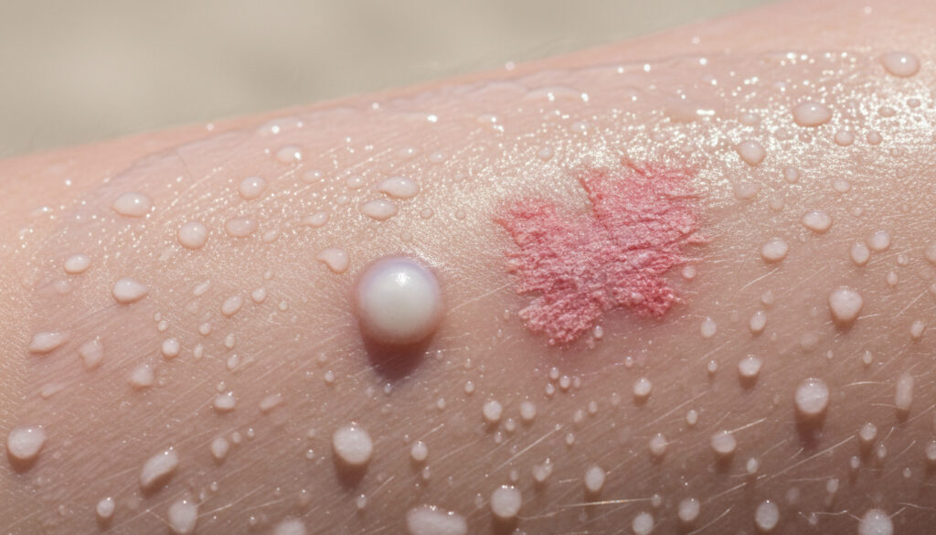
Clinical manifestations of basal cell carcinoma demonstrate significant heterogeneity depending on histological subtype, though certain morphological features remain consistent across presentations. The nodular subtype, accounting for the majority of cases, typically presents as pearly or waxy papules with raised, rolled borders and prominent telangiectasias. These lesions often display central ulceration or depression, earning the historical designation “rodent ulcer” due to their propensity for slow local tissue destruction.
What Does Basal Cell Carcinoma Look Like?
Superficial BCC presents as erythematous, scaly patches resembling eczema or psoriasis, frequently occurring on the trunk or extremities. Morpheaform (sclerosing) variants manifest as firm, scar-like, hypopigmented plaques with indistinct margins, posing particular diagnostic challenges due to their subtle appearance. Pigmented BCC contains melanin deposits, presenting as brown, blue, or black lesions that may simulate melanoma clinically.
Persistent lesions exhibiting pearly translucence, visible blood vessels, or cyclical bleeding and crusting without complete healing warrant immediate dermatological evaluation, particularly when located on sun-damaged skin.
Where Does BCC Typically Appear?
Anatomical distribution strongly correlates with cumulative sun exposure, with approximately 80% of lesions developing on the head and neck region. The nose, cheeks, and forehead represent particularly vulnerable sites. However, up to 20% of cases emerge on covered anatomical areas, including the trunk, genitals, or extremities, particularly in patients with predisposing genetic syndromes or history of radiation therapy. Penn Medicine emphasizes that any non-healing sore merits professional assessment regardless of location.
Causes and Risk Factors
The etiology of basal cell carcinoma involves multifactorial processes centered on cumulative genetic damage to keratinocyte progenitor cells. Ultraviolet radiation induces characteristic mutations in the Hedgehog signaling pathway, particularly affecting the PTCH1 tumor suppressor gene or the SMO proto-oncogene. This molecular disruption eliminates normal regulatory mechanisms controlling cellular proliferation and differentiation.
Ultraviolet Radiation and Environmental Exposures
Chronic sun exposure, particularly UVB wavelengths, constitutes the predominant carcinogenic stimulus, typically requiring a latency period of 15 to 20 years between initial DNA insult and clinical manifestation. The American Cancer Society identifies intermittent intense exposures resulting in sunburns, especially during childhood, as particularly deleterious. Indoor tanning devices emitting concentrated UVA contribute significantly to incidence rates among younger demographics.
Demographic and Genetic Predispositions
Phenotypic characteristics indicating reduced melanin protection—including Fitzpatrick skin types I and II, red or blonde hair, and light irides—correlate strongly with increased susceptibility. Male patients experience higher incidence rates, potentially reflecting occupational sun exposure patterns. Additional risk factors encompass prior radiation therapy, therapeutic immunosuppression following organ transplantation, chronic wound sites, and rare hereditary disorders such as Gorlin syndrome or xeroderma pigmentosum.
How Serious Is Basal Cell Skin Cancer?
Despite classification as a malignancy, basal cell carcinoma maintains the most favorable prognosis among cutaneous cancers, with survival rates approaching 100% following appropriate treatment. The biological behavior remains predominantly local, with metastatic dissemination occurring in fewer than 0.1% of cases, typically involving lymph nodes or distant organs only after decades of neglected growth.
Curability and Long-Term Outcomes
Standard surgical excision, including Mohs micrographic surgery for cosmetically sensitive or high-risk lesions, achieves five-year cure rates exceeding 95%. Recurrence rates vary by treatment modality and tumor characteristics, ranging from 5% for primary nodular lesions to 50% for previously treated morpheaform subtypes. The Cleveland Clinic notes that secondary BCCs develop in 40% of patients within five years, necessitating prolonged surveillance.
While basal cell carcinoma rarely proves fatal, melanoma claims approximately 7,000 lives annually in the United States despite representing fewer than 1% of total skin cancer diagnoses.
Growth Patterns and Local Invasion
The characteristic indolent growth pattern progresses over months to years, allowing ample opportunity for intervention. However, neglected lesions demonstrate locally aggressive behavior, eroding underlying cartilage, bone, and soft tissue structures. Perineural invasion, though uncommon, may facilitate extension along cranial nerves into the central nervous system, particularly in recurrent facial lesions.
Advanced basal cell carcinoma can cause significant disfigurement and functional impairment, occasionally requiring extensive reconstructive surgery when lesions invade critical anatomical structures.
From UV Exposure to Resolution: The Clinical Timeline
Understanding the chronological progression of basal cell carcinoma illuminates both its pathogenesis and the rationale for long-term management protocols. The disease trajectory spans decades, from initial carcinogenic insult to definitive treatment and subsequent surveillance.
- Cumulative UV Damage (15–20 years): Chronic ultraviolet radiation exposure induces progressive DNA damage in basaloid cells, particularly affecting the Hedgehog signaling pathway, without immediate clinical manifestation.
- Lesion Appearance (Months to Years): Clinically detectable lesions emerge slowly, often starting as small papules or patches that may bleed, crust, or ulcerate intermittently over substantial periods.
- Diagnostic Confirmation: Dermatological evaluation leads to biopsy—punch, shave, or excisional—providing histopathological confirmation of BCC subtype and informing treatment selection.
- Treatment Implementation: Surgical excision, Mohs surgery, or non-surgical modalities (topical imiquimod, photodynamic therapy) eliminate malignant tissue over several weeks, depending on lesion characteristics.
- Post-Treatment Surveillance (2–5 years): Regular dermatological examinations monitor for recurrence at the primary site and development of secondary BCCs, which occur in approximately 40% of patients within five years.
What We Know Versus What Remains Uncertain
Medical understanding of basal cell carcinoma rests on robust epidemiological and molecular evidence, though certain aspects of individual susceptibility and optimal management continue evolving.
| Established Medical Facts | Areas of Ongoing Investigation |
|---|---|
| UV radiation causes definitive DNA damage in basaloid cells | Specific threshold doses triggering malignancy in individual patients |
| Surgical excision cures >95% of primary lesions | Long-term comparative efficacy of emerging immunotherapies |
| BCC grows slowly over months to years | Triggers for aggressive subtypes in otherwise typical cases |
| Metastasis occurs in <0.1% of cases | Genetic modifiers determining immunosuppression-related risk |
| PTCH1 and SMO gene mutations drive most sporadic cases | Precise mechanisms of perineural invasion propagation |
Basal Cell Carcinoma Within the Skin Cancer Spectrum
Contextualizing BCC requires distinguishing its clinical behavior from malignant melanoma and squamous cell carcinoma. While melanoma originates from melanocytes and carries significant mortality risk through early metastasis, basal cell carcinoma remains confined locally, behaving more like a locally destructive benign process than a deadly cancer. Russian Losses in Ukraine – Verified Casualties and Equipment Data demonstrates how statistical tracking informs risk assessment, similar to dermatological surveillance protocols.
Public health initiatives increasingly emphasize prevention strategies applicable across all skin cancer types, including regular SPF 30+ application and avoidance of peak UV hours. The surveillance mechanisms for skin health parallel other monitoring systems, such as Met Office Weather Edinburgh – Hourly Forecast and Warnings, where consistent vigilance prevents adverse outcomes.
Medical Authority Perspectives
“Basal cell carcinoma is the most common form of skin cancer, with more than 3 million cases diagnosed in the United States each year. It rarely metastasizes and is highly curable when detected early.”
— American Academy of Dermatology
“The primary cause of BCC is ultraviolet (UV) radiation from the sun or tanning beds. People who use indoor tanning have a 29% increased risk of developing BCC before age 50.”
— Skin Cancer Foundation
Key Considerations for Basal Cell Carcinoma
Basal cell skin cancer, despite its prevalence, represents a highly manageable condition characterized by excellent cure rates and minimal mortality risk. Successful management hinges upon recognizing characteristic lesion features—pearly papules, non-healing sores, or scar-like plaques—and pursuing prompt dermatological evaluation. While treatment typically involves straightforward surgical excision, prevention through rigorous photoprotection remains paramount, particularly for fair-skinned individuals and those with significant sun exposure histories. Regular skin surveillance ensures early detection of both primary lesions and recurrences, maintaining the favorable prognosis that distinguishes this common malignancy from more aggressive skin cancers.
Frequently Asked Questions
Can basal cell carcinoma recur after successful treatment?
Recurrence rates range from 5% to 50% depending on tumor subtype, location, and treatment method. Patients have a 40% chance of developing a new, separate BCC within five years of initial diagnosis.
Does having BCC increase the risk of developing other cancers?
While BCC itself rarely metastasizes, patients with one skin cancer face increased risk of developing additional BCCs, squamous cell carcinomas, and melanoma, necessitating comprehensive dermatological surveillance.
Can basal cell carcinoma transform into melanoma?
BCC and melanoma originate from different cell types and BCC does not transform into melanoma. However, patients may develop both cancers independently, requiring careful examination of all suspicious lesions.
Is basal cell carcinoma contagious?
BCC results from genetic mutations caused by UV radiation exposure and cannot spread through physical contact, sharing personal items, or bodily fluids.
Do I need chemotherapy for basal cell skin cancer?
Standard chemotherapy proves ineffective against BCC. Advanced cases may require targeted oral medications called Hedgehog pathway inhibitors, such as vismodegib, which block specific molecular signals driving tumor growth.
What distinguishes Mohs surgery from standard excision?
Mohs micrographic surgery involves removing thin tissue layers and immediate microscopic examination, preserving healthy tissue while ensuring complete tumor removal—particularly valuable for facial lesions or recurrent tumors.
More related posts
 In the Night Garden Toys – Complete Buying Guide for Parents
 Tube Map with Elizabeth Line – Official TfL PDFs and Route Guide
 Good Series to Watch – Best TV Shows on Netflix, Prime & More
 Holly Valance – Biography, Net Worth and Political Shift in 2025
 Eva Longoria – Biography, Net Worth and Career in 2025
 Candyman (2021 Film) – Director, Cast, Box Office Facts
 Port Vale F.C. – League Two Profile, History and Fixtures
 A Level Periodic Table – Trends Across Periods and Groups
